We started the Paleontology module from a broad perspective, making sure to cover a brief history of life on earth as well as the history of evolutionary theory, including the work of such academic greats as Mayr, Simpson, and Dobzhansky. With such a solid base under our belts, we can now start learning mammalian anatomy!
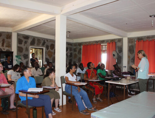
To be able to interpret fossil animal skeletons, it is helpful to begin with their modern descendants. We began the day with a classroom session, learning the proper terminology and understanding anatomical position.
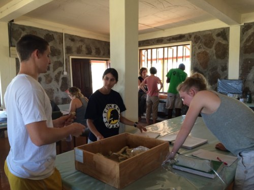
We spent the last two afternoons in the lab. The first day we focused on the different bones in the mammalian body and how to distinguish these bones of different kinds of animals. The second day we focused on differences in functional morphology.
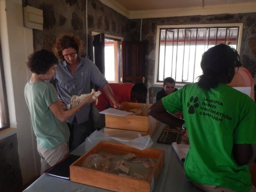
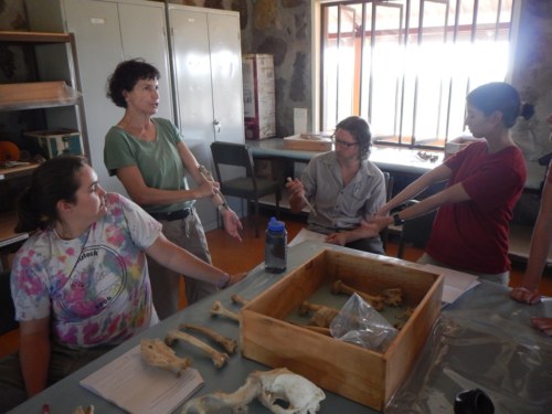
One of the most amazing aspects about learning paleontology at TBI is that, in addition to their vast fossil archives, there are a plethora of mammalian skeletons available for students to study! Thus, for our hands-on learning, we had a wide range of different mammalian specimen, including hyena, gerenuk, dik-dik, black-banded jackal, donkey, a juvenile baboon, and more!
Dr. Miller and Jon discuss prominent features seen on the warthog skull.
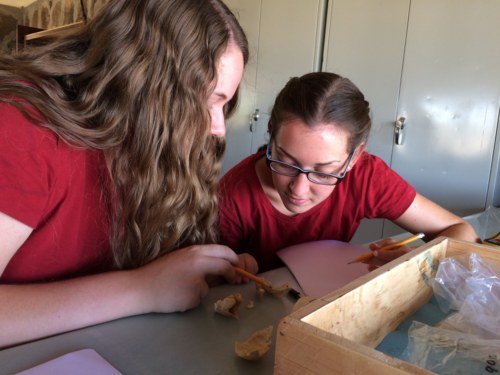
Carla meticulously sketches the fossil teeth.
Emily examines the zygomatic arch on the warthog skull as Dr. Miller teaches her quick tricks to identify it in the field.
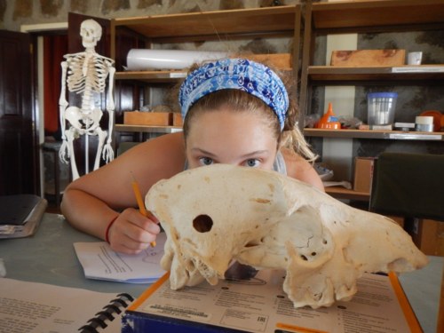
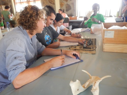
Morgan looks over her notes after completing a drawing of this warthog skull.
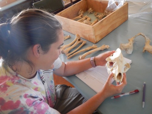
Kathryn closely studies this hyena skull.
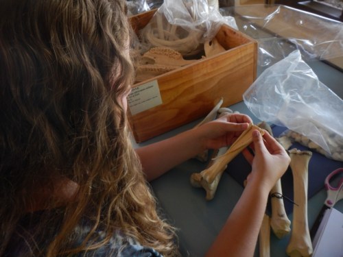
Morgan and Esther work together to learn key features on the scapula of this gerenuk specimen.
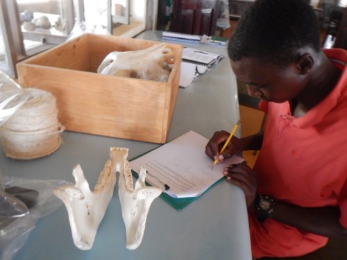
Yvette and Millie try to figure out the dental formula of this juvenile baboon by counting the teeth in its mandible
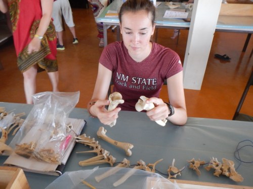
TA Mattia discusses with Carla how the radius and ulna of this hyena specimen articulate.
Dr. Miller explains to students how they can distinguish a left or right ulna by holding it in anatomical position.
Tobias and Jon discuss how to distinguish the left from right hyena humerus.
Functional morphology reflects how an animal’s skeletal morphology determines its adaptive niche. Thus, understanding the functional morphology of a living organism can help us understand behavioral aspects of fossil specimens and what kind of habitat they occupied. For example, we can extrapolate a lot about an organism from the type of teeth it has: animals with very high crowned teeth (equids, such as horses and zebras) tend to be grazers, eating large amounts of grasses and other tough vegetation. Grazing tends to wear teeth down rather rapidly, and as such, animals that spend their life eating a lot of grasses must have teeth that can last them their entire life. Other examples of functional morphology can be seen in the elongated vertebrae of the gerenuk; the powerful forearms of hyenas and warthogs; and the very wide zygomatic arch in hyenas.
Tobias investigates the mandible of a donkey, noting its specialized teeth.
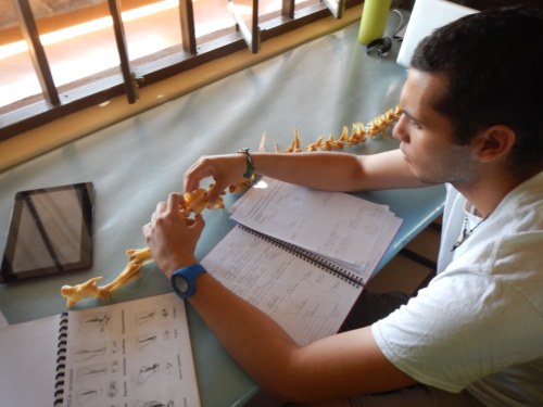
Max learns about the vertebrae of a gerenuk, pictured below. Note the elongated cervical vertebrae and how they correspond with the gerenuk’s elongated neck.
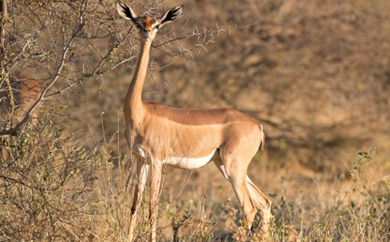
A gerenuk; note the long neck.
Yvette fits together the metatarsals of the donkey. The third is quite large and predominately makes up their foot, while the second and fourth metatarsals have become vestigial, shrinking to just slivers of bone.
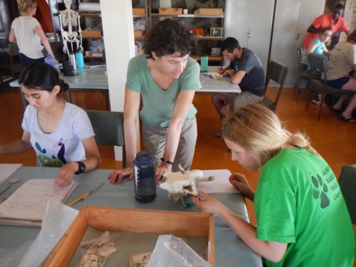
Danielle examines the humeri of a hyena (left) and warthog (right), as compared to the warthog femur (lying prone below hands). Hyenas and warthogs have very robust humeri, which can support large and powerful forearm musculature, advantageous for digging.
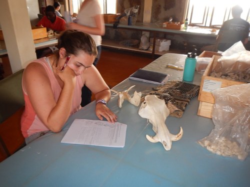
Morgan examines a hyena skull; the zygomatic arch (what look to be the wide cheekbones) is quite pronounced, and supports powerful jaw muscles, which the hyena uses to break through bone.
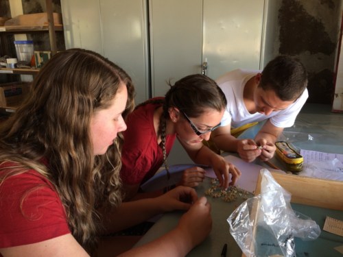
The students draw fossil teeth from several different specimen, paying particular attention to the differences in tooth morphology.
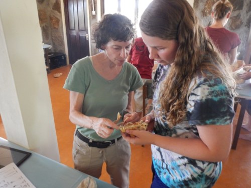
Dr. Miller and Yvette investigate the teeth of a black-banded jackal; Dr. Miller explains how the carnivore mouth and teeth have specialized for meat-eating.
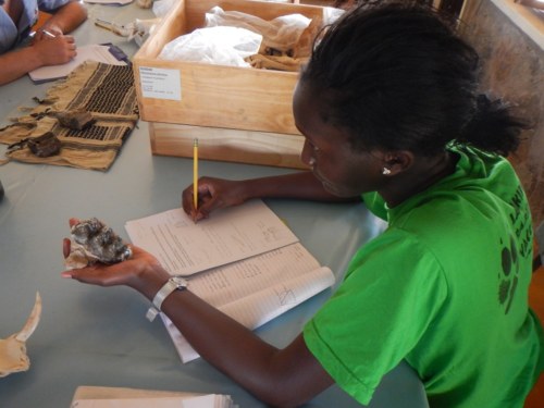
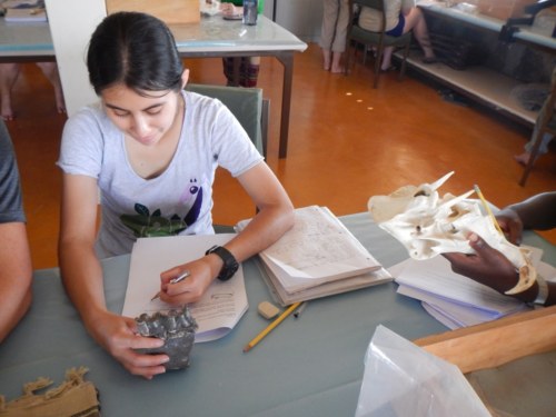
Esther draws a fossilized tooth; these teeth are thought to have belonged to an ancestral elephant relative from the Miocene.
Dr. Miller explains to students how to distinguish the astragalus of artiodactyls (even-toed ungulates) from perissodactyls (odd-toed ungulates).
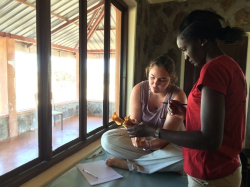
TA Mattia helps Millie and Yvette sort through the tiny vertebrae of a juvenile baboon.
Learning to identify mammalian anatomy and understanding functional morphology is paramount to interpreting fossils one may find out in the field! Over the next few days, the students will be spending more time in the lab looking at real fossils from the TBI archives, as well as learning about fossil preparation and proper field techniques!